The synovial fluid from patients with focal cartilage defects contains mesenchymal stem/stromal cells and macrophages with pro- and anti-inflammatory phenotypes

Abstract
Objective: The synovial fluid (SF) of patients with focal cartilage defects contains a population of poorly characterised cells that could have pathophysiological implications in early osteoarthritis and joint tissue repair. We have examined the cells within SF of such joints by determining their chondrogenic capacity following culture expansion and establishing the phenotypes of the macrophage subsets in non-cultured cells.
Design: Knee SF cells were obtained from 21 patients receiving cell therapy to treat a focal cartilage defect. Cell surface immunoprofiling for stem cell and putative chondrogenic markers, and the expression analysis of key chondrogenic and hypertrophic genes were conducted on culture-expanded SF cells prior to chondrogenesis. Flow cytometry was also used to determine the macrophage subsets in freshly isolated SF cells.
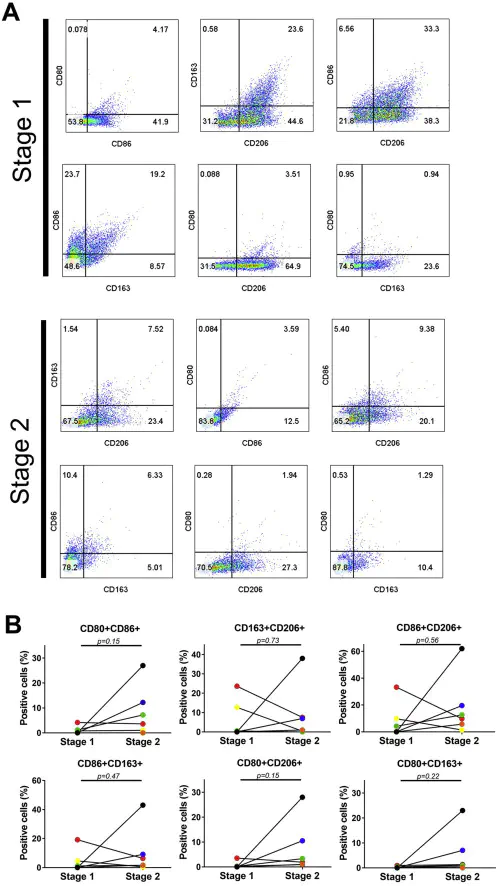
Results: Immunoprofiling revealed positivity for the monocyte/macrophage marker (CD14), the haematopoietic/endothelial cell marker (CD34) and mesenchymal stem/stromal cell markers (CD73, CD90, CD105) on culture expanded cells. We found strong correlations between the presence of CD14 and the vascular cell adhesion marker, CD106 (r = 0.81, p = 0.003). Collagen type II expression after culture expansion positively correlated with GAG production (r = 0.73, p = 0.006), whereas CD90 (r = −0.6, p = 0.03) and CD105 (r = −0.55, p = 0.04) immunopositivity were inversely related to GAG production. Freshly isolated SF cells were positive for both pro- (CD86) and anti-inflammatory markers (CD163 and CD206).
Conclusions: The cellular content of the SF from patients with focal cartilage injuries is comprised of a heterogeneous population of reparative and inflammatory cells. Additional investigations are needed to understand the role played by these cells in the attempted repair and inflammatory process in diseased joints.